Heart Scan or Coronary Artery Calcium (CAC) Score
- As many as 50% of the heart attack and deaths occur in people with no known history of coronary artery disease (CAD). Symptomatic heart disease has a long pre-clinical detectable phase ─silent heart disease that can be easily detected by elevated coronary artery calcium (CAC) score, popularly called a heart scan.1, 2 in other words, use of heart scan is helpful in saying if the process of atherosclerosis has started, and is helpful in choosing between lifestyle intervention versus lifestyle intervention plus statin therapy.
- The current practice of preventive cardiology demand matching the intensity of treatment to the severity of risk regardless of symptoms.3 The Framingham Risk Score (FRS) is considered the gold-standard for the determination of CAD risk and guides its management.
- The FRS significantly underestimates the CAD risk among those with family history of premature CAD, metabolic syndrome, as well as women, Asian Indians and other South Asians.4, 5 Most women remain at low risk by FRS until 70 years of age.6, 7
- Many vulnerable plaques that produce heart attack get calcified a late stage of the atherosclerotic process. Such calcified plaques can be quantified as CAC score using a CT Scan. CAC score is a powerful marker for the presence and extent of CAD regardless of symptoms.1, 2, 8 CAC score is also a strong determinant of heart attack in people with multiple risk factors.9
- Dyslipidemia (abnormal cholesterol and lipid levels) is the principal driver of CAC score.10 People with high LDL-C (low density lipoprotein cholesterol) are 7 times more likely to have elevated CAC score compared to those with low LDL-C (<70 mg/dl).11
- Nonoptimal levels of LDL-C and HDL-C (high density lipoprotein cholesterol) during young adulthood are independently associated with coronary atherosclerosis two decades later.12
- At any given age, non-whites have approximately half the CAC score of whites, and women have approximately half the calcium score of men.13
- A person with silent heart disease identified as CAC score of 300 to 400 or more is an indication for further diagnostic evaluation such as exercise testing. Conversely, symptomatic patients with CAC score in the low category can perhaps, at least temporarily, avoid expensive and invasive coronary angiography but not risk factor control.14
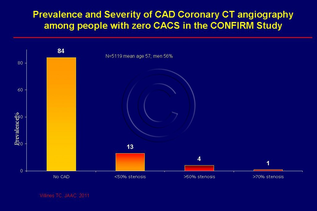
- In a large study involving 10,037 symptomatic patients (mean age 57 and 56% male) has recently confirmed the absence of significant obstructive coronary artery disease in people with calcium score zero which was found in 51% of subjects. Only one 1% of them had severe obstructive CAD. (Figure 113). Since 13% had mild CAD who could benefit from aggressive lipid lowering therapy, zero CACS is not a justification to deny appropriate guideline mandated treatment.32
Insights from MESA
- High hsCRP, as defined by JUPITER, was not associated with CAC in the absence of obesity. In contrast, obesity was associated with both measures of subclinical atherosclerosis independently of hsCRP status.15
- In The MESA study, per 1,000 person-years, rates of CAD events increased from 0.8 for patients with a CAC score of 0 to 20.2 for patients with a score greater than 100. Furthermore, 74% of all coronary heart disease events and 60% of all cardiovascular events occurred in the patients with a CAC score greater than 100. The five-year number needed to treat (NNT) to prevent one coronary heart disease event was 549 for patients with a CAC score of 0 and 23 for those with scores greater than 100.
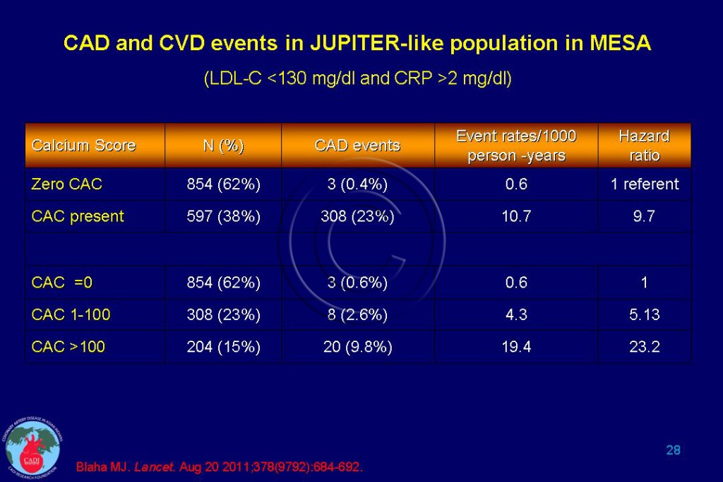
- Looking at patients with both low and high levels of hsCRP, the researchers found that CAC score — but not hsCRP — was associated with the risk of coronary heart disease events and overall cardiovascular events. CAC score could be used to target subgroups of patients who are expected to derive the most, and the least, absolute benefit from statin treatment. Compared to those with a CAC score of zero, those with less than 100 had 5-fold risk of heart attack, which increased to 23-fold in those who had a CAC score of more than 100 (Table 028).16
- A CAC score of 0 indicates an excellent prognosis with a 10 year coronary event rate (such as heart attack) of less than 2%.17 Those with CAC score of 1 to 10 have 2- 3-fold risk of coronary events compared to those with a CAC score of zero.18, 19 Increasing CAC score correspondingly increases the risk of a heart attack (Table 1A).
- Now that CAC scoring is so inexpensive (less than $100 in many centers), one can make the case of testing this in most patients at intermediate risk, and for the specific subset of low-risk patients with family history of CAD.20 The intermediate risk is generally defined as a 10-year risk between 10% and 20% (using FRS) but also includes young men and women with a CAD risk of 5-20% (low absolute risk but high relative risk).3, 20
|
Table 1 A. Risk of CAD with increasing Coronary artery calcium Score (CACS) 8, 14, 17, 18 |
||
| CAC Score | Extent of CAD | CAD risk |
| 0 | No CAD | 1 (referent) |
| 1-10 | Minimal CAD | 3-fold |
| 11-100 | Mild CAD | 4-fold |
| 101-300 | Moderate CAD | 6-8-fold |
| 301-1000 | Extensive CAD | 10-fold |
| >1000 | Very extensive CAD | 11-16-fold |
| * The risk is not much different for a CAC score between 300 and 400 | ||
- The risk threshold may be further reduced to 3-10% risk for Asian Indians.21 A history of Indian descent is tantamount to having a national history of CAD. An Indian man will reach intermediate risk status of 3% at age 35 without other risk factors or at age 30 with any one of the following: total cholesterol (TC) >200, LDL >160, HDL<45, blood pressure (BP) >130, smoking, or diabetes.21, 22
- An Indian woman will reach intermediate risk status of 3% at age 45 without other risk factors or age 40 with any one of the following: TC >280, LDL >160, HDL<45, BP >160, smoking, or diabetes.21
- The heightened risk of heart attacks in people with a family history of premature heart disease and/or metabolic syndrome is underestimated by FRS. These subjects would benefit from an estimation of the CAC score.23, 24
- People with clinically significant CAC score are reclassified as high- risk individuals requiring more aggressive risk factor management, particularly intensive statin therapy to achieve and maintain LDL <70 mg/dl.25, 26
- Aggressive treatment should be offered to the subset of individuals with low LDL cholesterol with measurable atherosclerosis as evidenced by a high CAC score could potentially reduce the overall healthcare cost, and preventing heart attack and avoiding expensive coronary interventions. 16
Radiation risk
- The benefit of CAC score determination needs to be weighed against the potential harmful effects of radiation and cancer. The mean effective radiation dose of a CT scan when using appropriate protocols is 1 mSv.27
- The estimated cancer risk of a single CT scan of a patient aged 55 years may result in a lifetime excess risk of 30 for men and 80 for women per million.28 But in appropriately selected patients the CT scans may identify 10,000-20,000 people with significant heart disease requiring intensive medical management and save at least 1,000-2,000 lives.29
- It is worth noting that the use of preventive measures such as statins, blood pressure control, and weight loss do not reduce the coronary calcium while reducing the risk of a heart attack. Since CAC score determination can improve cardiac management without incurring significant downstream medical cost, this test can serve as a gatekeeper for more expensive testings.30
CAC score Among Asian Indians
- Although CAC is highly correlated with coronary plaque burden and silent myocardial ischaemia in whites it fails to identify excess risk in Asian Indians. Researchers measured CAC in 2,369 asymptomatic men and women, aged 35 to 75 years, as part of the London Life Sciences Population (LOLIPOP) study. 518 subjects had CAC scores >100 Agatston units and of these 256 (49%) patients underwent myocardial perfusion scintigraphy (MPS). CAC scores were similar among Asian Indians and whites after adjustment for conventional risk factors. MPS abnormalities were seen in 56 (22%) subjects. 31
- Although presence of diabetes and increasing CAC were independent predictors for severity of silent myocardial ischemia, Asian Indians did not have MPS did not identify greater ischemia among Asian Indians compared with whites. This appears incongruent with almost 2-fold higher risk of CAD mortality observed in IA.3
FAQ
Q. Is there a difference in plaque between men and women?
Men have more calcified plaque, mixed plaque and more calcium on Agatston scoring but women had more noncalcified plaque. Calcified plaque is considered more advanced and possibly more stable, while mixed plaque may be a sign of intermediate progression and possibly more dangerous. Noncalcified plaques have been thought to denote early disease and can rupture, causing a heart attack or death.
Sources
1. Tunstall-Pedoe H, Morrison C, Woodward M, Fitzpatrick B, Watt G. Sex differences in myocardial infarction and coronary deaths in the Scottish MONICA population of Glasgow 1985 to 1991: Presentation, diagnosis, treatment, and 28-day case fatality of 3991 events in men and 1551 events in women. Circulation. 1996;93(11):1981-1992.
2. Ferket BS, Genders TS, Colkesen EB, et al. Systematic review of guidelines on imaging of asymptomatic coronary artery disease. J Am Coll Cardiol. Apr 12 2011;57(15):1591-1600.
3. Okwuosa T M, Greenland P, Ning H, et al. Distribution of Coronary Artery Calcium Scores by Framingham 10-Year Risk Strata in the MESA (Multi-Ethnic Study of Atherosclerosis) Potential Implications for Coronary Risk Assessment. J Am Coll Cardiol. May 3 2011;57(18):1838-1845.
4. Blumenthal RS, Hasan RK. “Actually, it is more of a guideline than a rule”. J Am Coll Cardiol. Apr 12 2011;57(15):1601-1603.
5. Enas EA, Singh V, Munjal YP, Bhandari S, Yadave RD, Manchanda SC. Reducing the burden of coronary artery disease in India: challenges and opportunities. Indian Heart J. Mar-Apr 2008;60(2):161-175.
6. Ford ES, Giles WH, Mokdad AH. The distribution of 10-Year risk for coronary heart disease among US adults: findings from the National Health and Nutrition Examination Survey III. J Am Coll Cardiol. May 19 2004;43(10):1791-1796.
7. Pencina MJ, D’Agostino RB, Sr., Larson MG, Massaro JM, Vasan RS. Predicting the 30-year risk of cardiovascular disease: the framingham heart study. Circulation. Jun 23 2009;119(24):3078-3084.
8. Detrano R, Guerci AD, Carr JJ, et al. Coronary calcium as a predictor of coronary events in four racial or ethnic groups. N Engl J Med. Mar 27 2008;358(13):1336-1345.
9. Blankstein R, Budoff MJ, Shaw LJ, et al. Predictors of Coronary Heart Disease Events Among Asymptomatic Persons With Low Low-Density Lipoprotein Cholesterol MESA (Multi-Ethnic Study of Atherosclerosis). J Am Coll Cardiol. Jul 19 2011;58(4):364-374.
10. Paramsothy P, Knopp RH, Bertoni AG, et al. Association of combinations of lipid parameters with carotid intima-media thickness and coronary artery calcium in the MESA (Multi-Ethnic Study of Atherosclerosis). J Am Coll Cardiol. Sep 21 2010;56(13):1034-1041.
11. Newman TB, Pletcher MJ. Coronary calcium screening. N Engl J Med. Dec 17 2009;361(25):2491; author reply 2491-2492.
12. Pletcher MJ, Bibbins-Domingo K, Liu K, et al. Nonoptimal lipids commonly present in young adults and coronary calcium later in life: the CARDIA (Coronary Artery Risk Development in Young Adults) study. Ann Intern Med. Aug 3 2010;153(3):137-146.
13. Nasir K, Shaw LJ, Liu ST, et al. Ethnic differences in the prognostic value of coronary artery calcification for all-cause mortality. J Am Coll Cardiol. Sep 4 2007;50(10):953-960.
14. Dendukuri N, Chiu K, Brophy JM. Validity of electron beam computed tomography for coronary artery disease: asystematic review and meta-analysis. BMC Med. 2007;5:35.
15. Blaha MJ, Rivera JJ, Budoff MJ, et al. Association Between Obesity, High-Sensitivity C-Reactive Protein >=2 mg/L, and Subclinical Atherosclerosis: Implications of JUPITER from the Multi-Ethnic Study of Atherosclerosis. Arterioscler Thromb Vasc Biol. Jun 2011;31(6):1430-1438.
16. Blaha MJ, Budoff MJ, DeFilippis AP, et al. Associations between C-reactive protein, coronary artery calcium, and cardiovascular events: implications for the JUPITER population from MESA, a population-based cohort study. Lancet. Aug 20 2011;378(9792):684-692.
17. Erbel R, Mohlenkamp S, Moebus S, et al. Coronary risk stratification, discrimination, and reclassification improvement based on quantification of subclinical coronary atherosclerosis: the Heinz Nixdorf Recall study. J Am Coll Cardiol. Oct 19 2010;56(17):1397-1406.
18. Budoff MJ, McClelland RL, Nasir K, et al. Cardiovascular events with absent or minimal coronary calcification: the Multi-Ethnic Study of Atherosclerosis (MESA). Am Heart J. Oct 2009;158(4):554-561.
19. Blaha M, Budoff MJ, Shaw LJ, et al. Absence of coronary artery calcification and all-cause mortality. JACC Cardiovasc Imaging. Jun 2009;2(6):692-700.
20. Taylor AJ, Cerqueira M, Hodgson JM, et al. ACCF/SCCT/ACR/AHA/ASE/ASNC/NASCI/SCAI/SCMR 2010 appropriate use criteria for cardiac computed tomography: a report of the American College of Cardiology Foundation Appropriate Use Criteria Task Force, the Society of Cardiovascular Computed Tomography, the American College of Radiology, the American Heart Association, the American Society of Echocardiography, the American Society of Nuclear Cardiology, the North American Society for Cardiovascular Imaging, the Society for Cardiovascular Angiography and Interventions, and the Society for Cardiovascular Magnetic Resonance. J Am Coll Cardiol. Nov 23 2010;56(22):1864-1894.
21. Enas EA, Singh V, Gupta R, Patel R, et al. Recommendations of the Second Indo-US Health Summit for the prevention and control of cardiovascular disease among Asian Indians. Indian Heart J. 2009;61:265-74.
22. Greenland P, Alpert JS, Beller GA, et al. 2010 ACCF/AHA guideline for assessment of cardiovascular risk in asymptomatic adults: a report of the American College of Cardiology Foundation/American Heart Association Task Force on Practice Guidelines. J Am Coll Cardiol. Dec 14 2010;56(25):e50-103.
23. Lakoski SG, Greenland P, Wong ND, et al. Coronary artery calcium scores and risk for cardiovascular events in women classified as “low risk” based on Framingham risk score: the multi-ethnic study of atherosclerosis (MESA). Arch Intern Med. Dec 10 2007;167(22):2437-2442.
24. Scheuner MT, Setodji CM, Pankow JS, Blumenthal RS, Keeler E. General Cardiovascular Risk Profile identifies advanced coronary artery calcium and is improved by family history: the multiethnic study of atherosclerosis. Circ Cardiovasc Genet. Feb 1 2010;3(1):97-105.
25. Nasir K, McClelland RL, Blumenthal RS, et al. Coronary artery calcium in relation to initiation and continuation of cardiovascular preventive medications: The Multi-Ethnic Study of Atherosclerosis (MESA). Circ Cardiovasc Qual Outcomes. May 2010;3(3):228-235.
26. Polonsky TS, McClelland RL, Jorgensen NW, et al. Coronary artery calcium score and risk classification for coronary heart disease prediction. JAMA. Apr 28 2010;303(16):1610-1616.
27. Gerber TC, Gibbons RJ. Weighing the risks and benefits of cardiac imaging with ionizing radiation. JACC Cardiovasc Imaging. May 2010;3(5):528-535.
28. Kim K P, Einstein AJ, Berrington de Gonzalez A. Coronary artery calcification screening: estimated radiation dose and cancer risk. Arch Intern Med. Jul 13 2009;169(13):1188-1194.
29. Gerber TC, Carr JJ, Arai AE, et al. Ionizing radiation in cardiac imaging: a science advisory from the American Heart Association Committee on Cardiac Imaging of the Council on Clinical Cardiology and Committee on Cardiovascular Imaging and Intervention of the Council on Cardiovascular Radiology and Intervention. Circulation. Feb 24 2009;119(7):1056-1065.
30. Rozanski A, Gransar H, Shaw LJ, et al. Impact of coronary artery calcium scanning on coronary risk factors and downstream testing the EISNER (Early Identification of Subclinical Atherosclerosis by Noninvasive Imaging Research) prospective randomized trial. J Am Coll Cardiol. Apr 12 2011;57(15):1622-1632.
31. Jain P, Kooner JS, Raval U, Lahiri A. Prevalence of coronary artery calcium scores and silent myocardial ischaemia was similar in Indian Asians and European whites in a cross-sectional study of asymptomatic subjects from a U.K. population (LOLIPOP-IPC). J Nucl Cardiol. May 2011;18(3):435-442.
32. Villines TC, Hulten EA, Shaw LJ, et al. Prevalence and severity of coronary artery disease and adverse events among symptomatic patients with coronary artery calcification scores of zero undergoing coronary computed tomography angiography: results from the CONFIRM (Coronary CT Angiography Evaluation for Clinical Outcomes: An International Multicenter) registry. J Am Coll Cardiol. Dec 6 2011;58(24):2533-2540.


